- Anatomical terminology
- Skeletal system
- Skull
- Skeleton of trunk
- Skeleton of upper limb
- Skeleton of lower limb
- Joints
- Muscles
- Heart
- Blood vessels
- Nervous system
- Respiratory system
- Digestive system
- Lymphatic system
- Female reproductive system
- Male reproductive system
- Endocrine glands
- Eye
- Ear
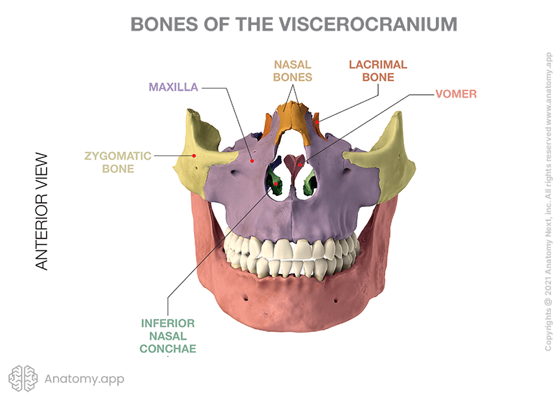
Viscerocranium
The viscerocranium (Latin: viscerocranium) is the part of the skull situated anterior to the neurocranium, and is made up of fourteen bones. It forms the facial skeleton and jaw, and acts as the framework for the soft tissues of the face.
The 14 bones forming the viscerocranium are the lacrimal bones (2), nasal bones (2), zygomatic bones (2), palatine bones (2), maxillary bones or maxillae (2), inferior nasal concha, vomer, and the mandible.
In some cases, the unpaired hyoid bone is also classified as part of the viscerocranium, although located in the upper neck region. It is connected to the skull with the help of ligaments. However, it is a separate structure from the skull.
Bones of viscerocranium
There are six paired and two unpaired bones forming the viscerocranium. Paired bones are found symmetrically located at opposite sides of the skull. The paired bones of the viscerocranium are the following:
- Lacrimal bone
- Nasal bone
- Zygomatic bone
- Palatine bone
- Maxilla
- Inferior nasal concha
Unpaired bones are symmetrical single bones that are located along the midline of the skull. And the unpaired bones of the viscerocranium are:
- Vomer
- Mandible
Paired bones of viscerocranium
Lacrimal bone
The lacrimal bones are very thin and fragile rectangular-shaped bones. They form the medial wall of the orbit and a small portion of the nasal cavity where they articulate with the ethmoid bone, maxilla, and inferior nasal concha.
Superiorly, the lacrimal bones articulate with the frontal bone. Inferior to this, the lacrimal bones articulate with the frontal process of the maxilla. In this area, in the lacrimal fossa of the lacrimal bone, the lacrimal sac is located.
Nasal bone
The nasal bones are located at the midline of the frontal viscerocranium. They form the nose bridge and articulate at the middle of the bridge with the opposing nasal bone. Inferior parts of these bones form the superior margin of the nasal aperture and attach to the lateral and septal nasal cartilages.
The nasal bones are in contact with three other bones of the skull. Superiorly, the nasal bones articulate with the nasal part of the frontal bone. Laterally, they articulate with the frontal process of the maxilla. And the nasal bones articulate with the ethmoid bone posteriorly.
Zygomatic bone
The zygomatic bone acts as a scaffold for the cheek, forms the zygomatic arch, and provides attachment sites for facial muscles. The zygomatic bone has several parts: a body and three processes - the frontal, temporal, and maxillary processes.
The frontal process and maxillary process both are involved in forming the orbits. They articulate with the frontal and maxillary bones. The zygomatic bone's temporal process forms the zygomatic arch by articulating with the zygomatic process of the temporal bone.
Palatine bone
The palatine bones are L-shaped bones that form part of the posterior nasal cavity, hard palate, and a small part of the orbit. Each palatine bone is made up of a perpendicular and a horizontal plate. The perpendicular plate forms an articulation with the sphenoid bone and maxilla.
The horizontal plates of the palatine bones are fused at the midline forming the hard palate that separates the nasal cavity and the roof of the mouth. At the point of fusion of the horizontal plates, the palatine bones articulate with the vomer.
Maxilla
The maxillae, also known as maxillary bones, are two viscerocranial bones that fuse into a single bone called the maxilla. The maxilla forms the upper jaw and contributes to the hard palate, orbit, nasal bridge, and infratemporal fossa. It is also the most protruding fixed bone of the facial skeleton. The maxilla contains a pair of paranasal sinuses - the maxillary sinuses.
The main parts of the maxilla are the body and four processes. These are the frontal, zygomatic, palatine, and alveolar processes. The frontal process articulates superiorly with the nasal, lacrimal, frontal, and ethmoid bones. The zygomatic process articulates with the zygomatic bone. The palatine process contributes to formation of the hard palate. And the alveolar process supports all the upper (maxillary) teeth.
Inferior nasal concha
The inferior nasal concha is a curved bone located within the lateral wall of the nasal cavity. The inferior nasal concha articulates with the palatine and lacrimal bones, the ethmoid, and the maxilla.
Unpaired bones of viscerocranium
Vomer
The vomer is a plow-shaped bone that forms the posteroinferior part of the nasal septum. Superiorly, the vomer articulates with the ethmoid bone and sphenoid bone. Inferiorly, the vomer articulates with the maxilla and palatine bones. Anteriorly, the vomer articulates with the septal cartilage. The posterior border of the vomer is free.
Mandible
The mandible is a horseshoe-shaped bone that forms the lower jaw. It is the site of attachment for all primary muscles of mastication. The mandible is detached from the skull but articulates with it at the temporomandibular joint. The mandible consists of a body, rami (2), coronoid processes (2), condylar processes (2), and alveolar process.
The condylar process articulates with the temporal bone in the temporomandibular joint. The coronoid process is a bony projection anterior to the condylar process. It is the attachment site for the temporalis and masseter muscles. The alveolar process of the mandible supports the lower (mandibular) teeth.
References:
- Drake, R. L., Vogl, A. W., & Mitchell, A. W. M. (2019). Gray’s Anatomy for Students: With Student Consult Online Access (4th ed.). Elsevier.
- Norton, N. S. (2016). Osteology. In Netter's Head and Neck Anatomy for Dentistry (3rd ed., pp. 25–64). Elsevier.
Anatomy.app
Contact information
- For questions regarding business inquiries. Please contact:
- info@anatomy.app