- Anatomical terminology
- Skeletal system
- Skull
- Skeleton of trunk
- Skeleton of upper limb
- Skeleton of lower limb
- Joints
- Muscles
- Heart
- Blood vessels
- Nervous system
- Respiratory system
- Digestive system
- Lymphatic system
- Female reproductive system
- Male reproductive system
- Endocrine glands
- Eye
- Ear
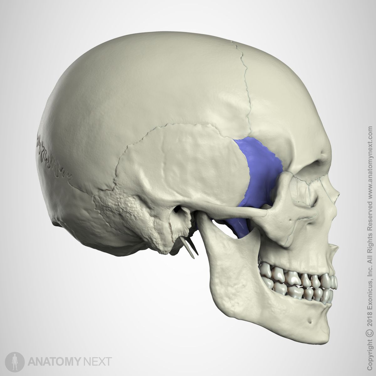
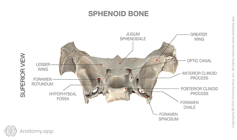
Sphenoid bone
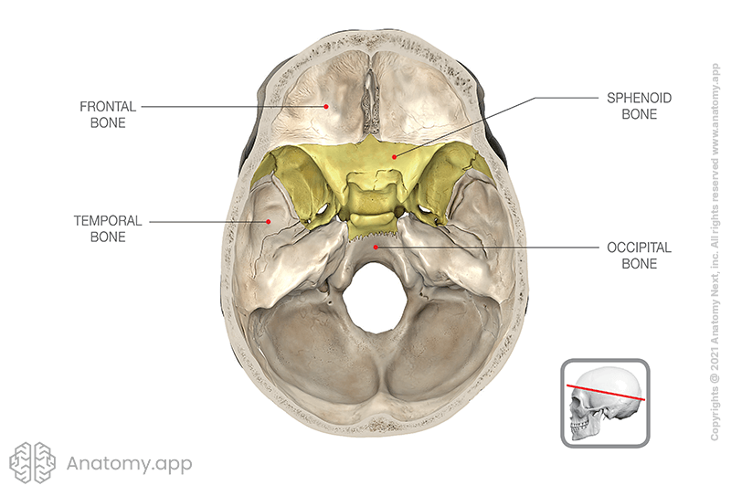
The sphenoid bone (also called the sphenoid, sphenoidal bone, Latin: os sphenoidale) is a single bone that lies in the base of the skull between the frontal, temporal and occipital bones.
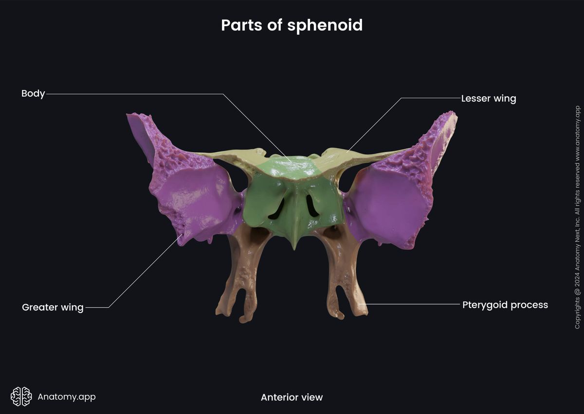
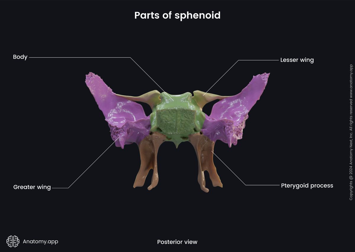
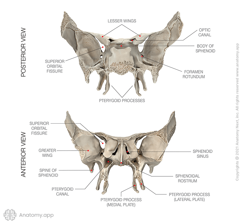
The sphenoid bone has a central body, and paired greater wings and lesser wings that extend laterally from the body of the sphenoid. It also has two pterygoid processes descending from the junction of the body and greater wings.

Parts of sphenoid bone
The main parts of the sphenoid bone are as mentioned before:
- Body
- Lesser wing (2)
- Greater wing (2)
- Pterygoid process (2)
Body of sphenoid
The body of the sphenoid (or sphenoid body) is the central portion of the sphenoid bone located between the wings and processes of the bone. The sphenoid body houses the sphenoidal sinuses. The body has six surfaces: posterior, anterior, inferior, superior, and two lateral surfaces.
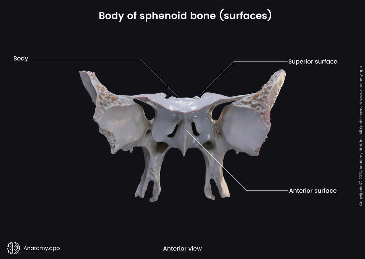
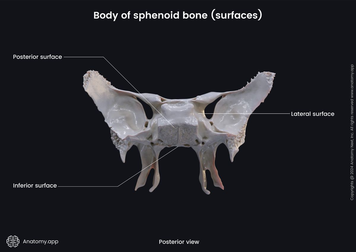
The posterior surface of the sphenoid body is the part that joins with the basilar part of the occipital bone. The anterior surface of the body presents the opening of the sphenoidal sinus (2) and septum of the sphenoidal sinuses.
The opening of the sphenoidal sinus is an aperture that opens anteriorly into the nasal cavity, specifically, into a small space called the spheno-ethmoidal recess. As mentioned before, the sphenoidal sinus is a paired air-filled cavity located within the body of the sphenoid. And the septum of the sphenoidal sinuses is a bony partition separating the two sinuses.
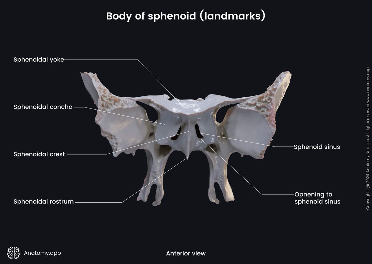
The inferior surface of the sphenoid body features in the middle line a triangular spine called the sphenoidal rostrum. It fits into a deep fissure between the alae of the vomer. And the superior surface of the sphenoid body features the following landmarks:
- Chiasmatic groove
- Sella turcica with:
- Hypophysial fossa
- Tuberculum sellae
- Dorsum sellae
The chiasmatic groove (also known as optic groove) is a transverse groove between the right and left optic canals. It is continuous with the optic openings, located just anterior to the sella turcica. Within the chiasmatic groove lies the optic chiasm - junction of the two optic nerves (CN II).
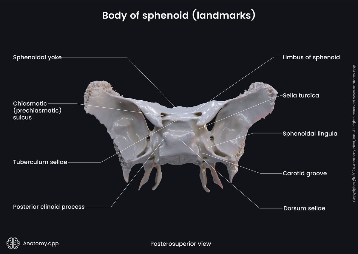
The sella turcica (or turkish saddle) is a saddle-shaped depression on the body of the sphenoid. It lies above the sphenoidal sinus and contains the hypophysis (pituitary gland). The sella turcica features such structures as the hypohysial fossa, tuberculum sellae and dorsum sellae.
The hypophysial fossa (or pituitary fossa) is a depression on the superior surface of the sphenoid body occupied by the hypophysis. The tuberculum sellae is a small process in front of the hypophysial fossa. And the dorsum sellae is the posterior wall of the hypophysial fossa.
Each lateral surface of the sphenoid body unites with the greater wing and the pterygoid process of the sphenoid. This surface also has an important feature, which is called the carotid groove. The carotid groove is a longitudinal groove that houses the internal carotid artery and the cavernous sinus.
Greater wings of sphenoid
The greater wings of the sphenoid are the larger of the two lateral, wing-like extensions of the sphenoid body. The greater wings curve upwards, laterally and backwards to form a prominent concavity or fossa in the middle of the cranial floor.
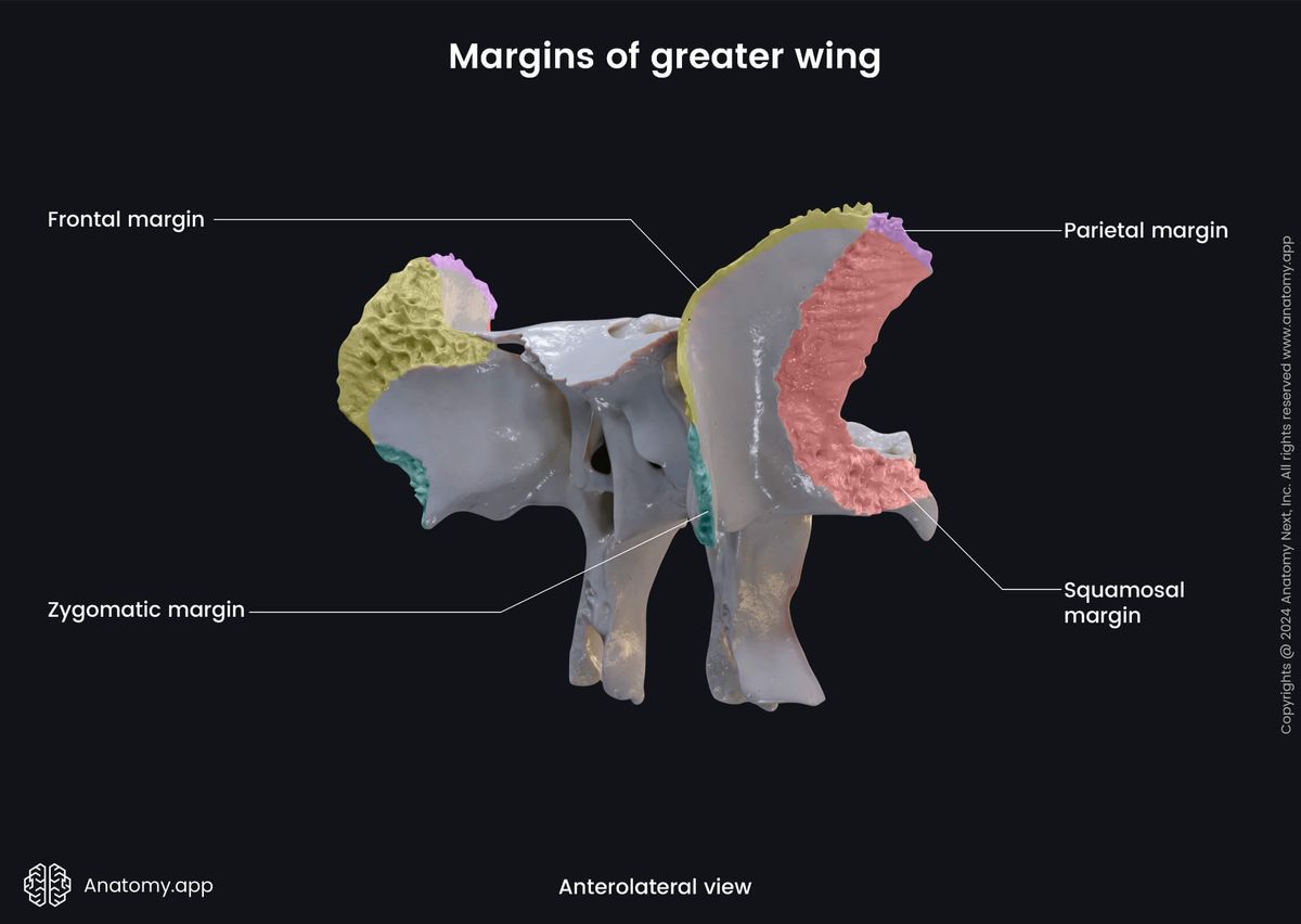
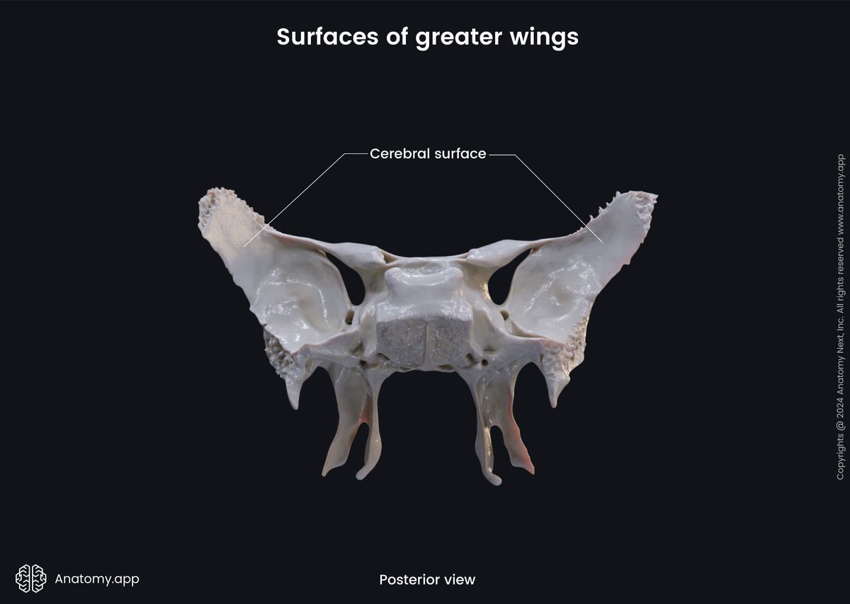
Each greater wing of the sphenoid has five surface (cerebral surface, lateral surface (composed of two parts - temporal and infratemporal surfaces), maxillary surface, and orbital surface). Besides the mentioned surfaces, the greater wings has the following four margins:
- Zygomatic margin - border that articulates with the zygomatic bone;
- Frontal margin - border that articulates with the frontal bone;
- Parietal margin - border that connects with the parietal bone;
- Squamosal margin - border that connects with the temporal bone.
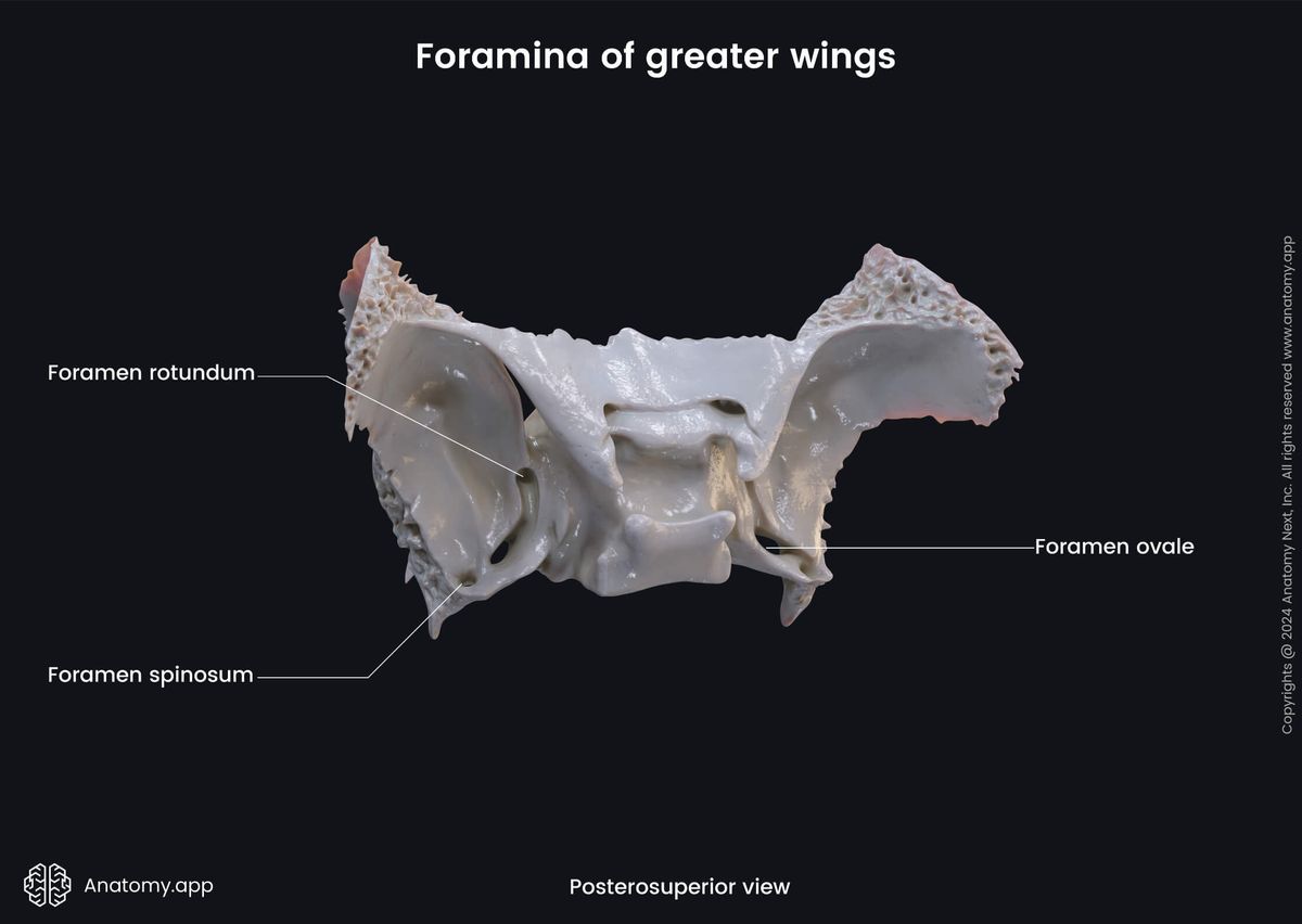
The cerebral surface of the greater wing forms the anterior part of the middle cranial fossa. This surface has the following features:
- Foramen rotundum
- Foramen ovale
- Foramen spinosum
- Impressions of cerebral gyri
- Arterial grooves
The foramen rotundum is a circular hole in the greater wing of the sphenoid connecting the middle cranial fossa to the pterygopalatine fossa. It is a passage for the maxillary branch of the trigeminal nerve (CN V2).
The foramen ovale is an opening that connects the middle cranial fossa to the external surface of the skull. It transmits several vessels and nerves, including the mandibular nerve (CN V3), the motor root of the trigeminal nerve (CN V), and the lesser petrosal nerve (a component of the glossopharyngeal nerve, CN IX).
The foramen spinosum is a round, small opening in the greater wing located posterolateral to the foramen ovale. It connects the middle cranial fossa to the external surface of the skull. The foramen spinosum transmits the middle meningeal artery and vein, and the meningeal branch of the mandibular nerve (CN V3).
The impressions of cerebral gyri (also called the digital impressions) are flat indentations on the inner surface of the skull corresponding to the cerebral gyri which produce them. Also, the inner surface features arterial grooves that are produced by pressure from arteries, primarily the middle meningeal artery and its branches.
The lateral surface of the greater wing of the sphenoid is convex and divided into two parts (temporal and infratemporal surfaces) by a transverse bony ridge called the infratemporal crest. The lateral surface of the greater wing is pierced by the foramen ovale and foramen spinosum. It also features the downward projecting sphenoidal spine.
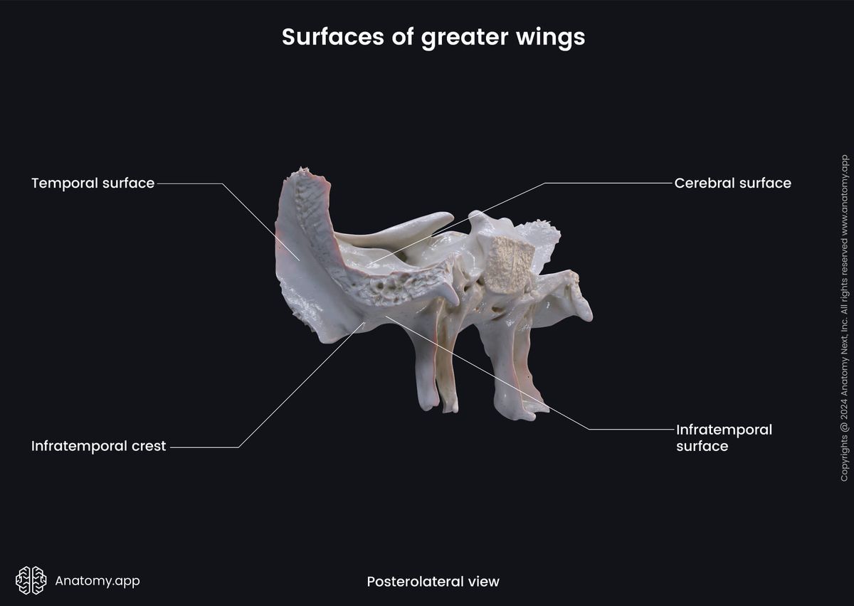
The upper vertical part of the lateral surface is the temporal surface. It is the attachment site for the temporalis muscle. The horizontally-oriented lower portion is known as the infratemporal surface. It is directed downwards and together with the infratemporal crest is the attachment site of the upper fibers of the lateral pterygoid muscle.
The maxillary surface of the greater wing faces anteriorly and forms the posterior border of the pterygopalatine fossa. This surface is pierced by the foramen rotundum. And the orbital surface of the greater wing faces anteromedially. It forms the posterior part of the lateral wall of the orbit.
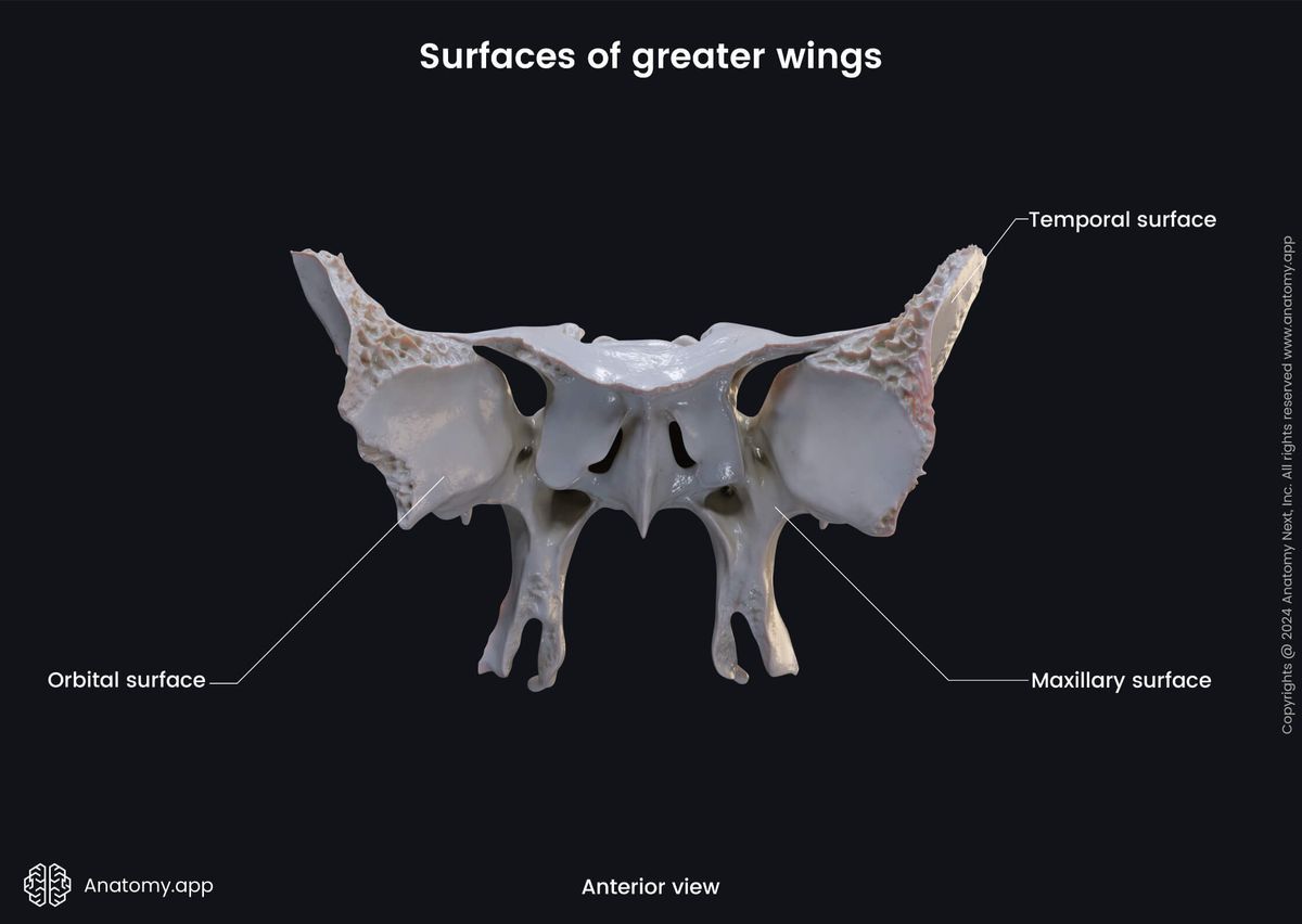
The upper edge of the orbital surface of the greater wing articulates with the orbital plate of the frontal bone, while the lateral margin articulates with the zygomatic bone. The inferior margin of the orbital surface forms the posterolateral margin of the inferior orbital fissure, while its medial margin forms the inferolateral edge of the superior orbital fissure.
Lesser wings of sphenoid
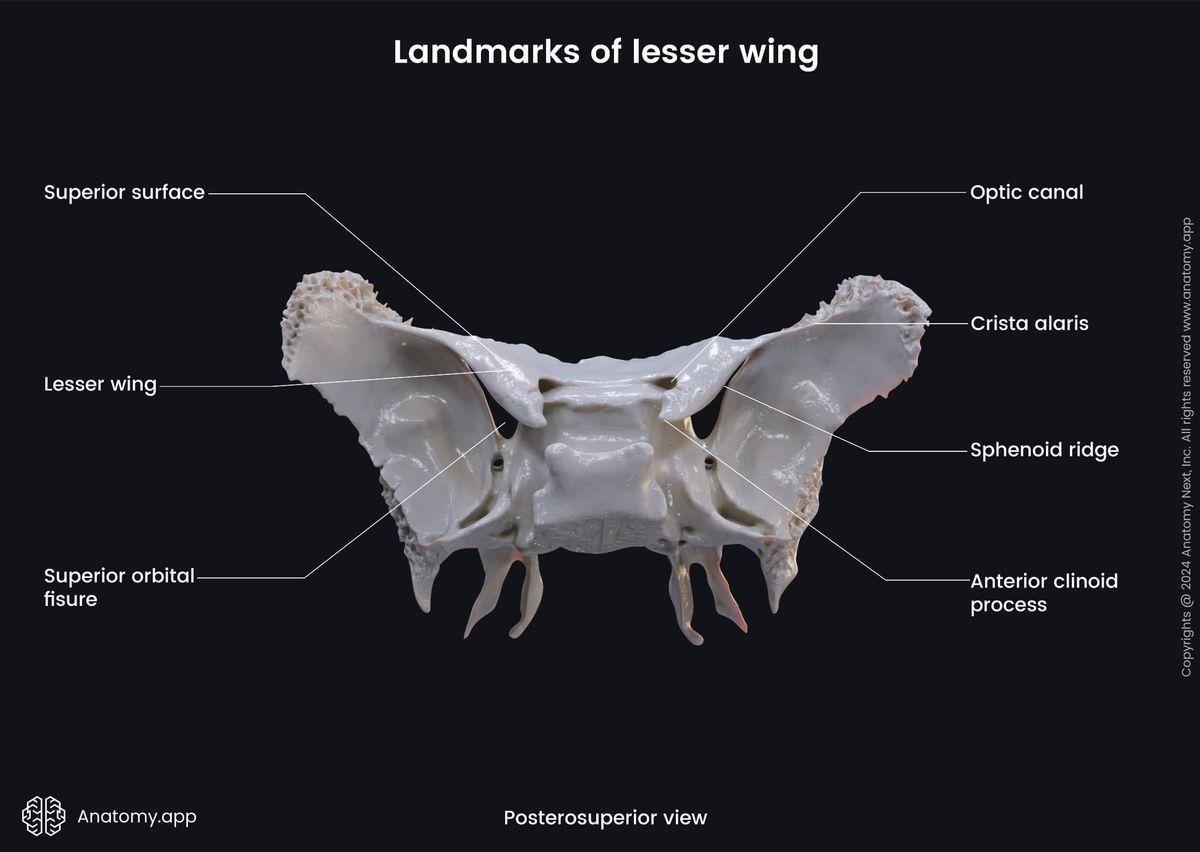
The lesser wings of the sphenoid are the smaller of two lateral wing-like extensions of the sphenoid body. They are flattened, triangular in shape, and located above and anterior to the greater wings. Each lesser wing of the sphenoid has two surfaces (cerebral and orbital), and features the following structures:
- Optic canal
- Anterior clinoid process
The optic canal is a passage connecting the orbit to the middle cranial fossa. It transmits the optic nerve (CN II) and the ophthalmic artery. The anterior clinoid process is a cone-like process of the sphenoid on either side of the anterior part of the hypophysial fossa.
Also, the lesser wing of the sphenoid contributes to forming the superior orbital fissure. It is the gap between the greater and lesser wings of the sphenoid bone, bordered medially by the sphenoid body, and closed at its anterior extremity by the frontal bone.
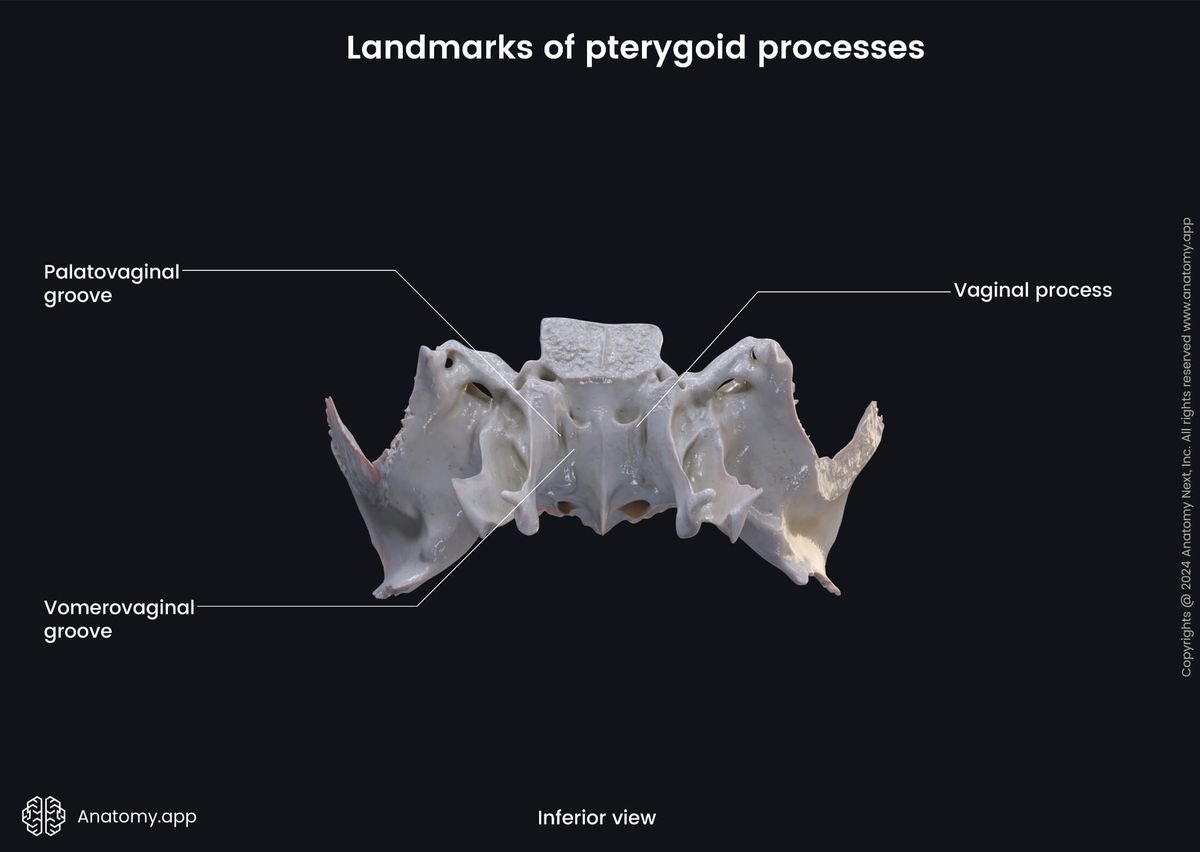
Pterygoid processes
The pterygoid process is a paired posteroinferior projection of the sphenoid bone. Each process bifurcates into a medial and a lateral plate. Each pterygoid process also features the pterygoid fossa and pterygoid canal.
The lateral plate of the pterygoid process is the lateral division of the process where the pterygoid muscles attach. The lateral pterygoid muscle to the lateral aspect, and the medial pterygoid muscle - to the medial aspect of the lateral plate.
The medial plate is the medial division of the pterygoid process. The superior pharyngeal constrictor muscle attaches to the inferior end of this plate.
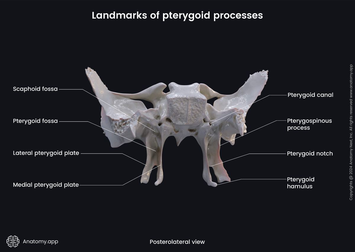
Between the lateral and medial plates of each pterygoid process there is a space called the pterygoid fossa. It is the attachment site of the medial pterygoid muscle.
The pterygoid canal is a bony passage that extends anteriorly in the base of the pterygoid proces. This canal transmits the greater and deep petrosal nerves from the middle cranial fossa to the pterygoid ganglion in the pterygopalatine fossa.
Anatomy.app
Contact information
- For questions regarding business inquiries. Please contact:
- info@anatomy.app