- Anatomical terminology
- Skeletal system
- Joints
- Muscles
- Heart
- Blood vessels
- Nervous system
- Respiratory system
- Digestive system
- Lymphatic system
- Female reproductive system
- Male reproductive system
- Endocrine glands
- Eye
- Ear
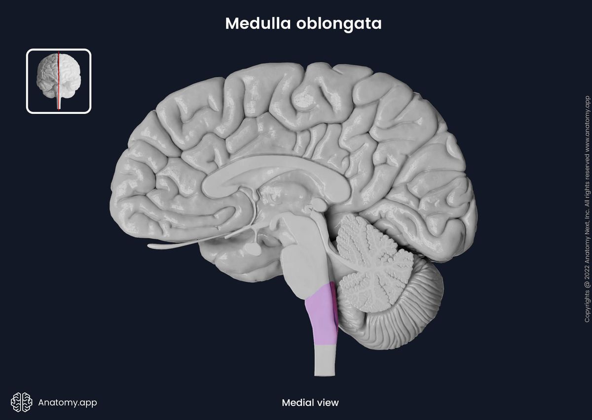
Medulla oblongata
The medulla oblongata (Latin: medulla oblongata) is the most caudal portion of the brainstem located in the posterior cranial fossa. The upper aspect of the medulla oblongata connects to the pons, while its lower part continues as the spinal cord. Besides being a conduit for fibers running between the spinal cord and higher brain regions, the medulla oblongata also contains control centers for essential involuntary functions such as regulation of blood pressure and breathing.
Anatomically, the medulla oblongata has external and internal features. Externally, the medulla oblongata has two surfaces: ventral (anterior) and dorsal (posterior). The ventral surface of the medulla faces the clivus of the occipital bone. The upper rear part of the medulla oblongata forms the lower portion of the fourth ventricle. Four pairs of cranial nerves emerge on the ventral surface of the medulla oblongata: glossopharyngeal (CN IX), vagus (CN X), accessory (CN XI), and hypoglossal (CN XII) nerves.
The dorsal surface of the medulla oblongata forms the inferior part of the rhomboid fossa and is connected to the cerebellum by the inferior cerebellar peduncles. Moreover, this surface also creates the inferior aspect of the fourth ventricle’s floor and the obex - the most caudal part of the fourth ventricle.
When looked at in cross-section, the medulla oblongata can be subdivided into three parts: basis, tegmentum, and tectum. The basis houses the pyramidal decussation, the tegmentum contains several nuclei and tracts, and the tectum consists of the lower aspect of the fourth ventricle covered by the inferior medullary velum.
The internal anatomy of the medulla oblongata is very complex as it contains many nuclei (clusters of neuron cell bodies) and tracts (consisting of nerve fibers). To better understand these structures and their relations, the medulla oblongata is typically examined in three cross-section levels: at the pyramidal decussation level, sensory decussation level, and the level of the olives.
Medulla oblongata external anatomy
The medulla oblongata appears cone-shaped, and it slightly decreases in width as it descends. It is 2.5 - 3.5 cm long and 2 - 2.5 cm broad at its widest part. The medulla oblongata is separated from the pons above by the inferior pontine sulcus that marks the pontomedullary junction. Inferiorly, the medulla oblongata continues as the spinal cord. The external anatomy of the medulla oblongata includes structures found on its two surfaces: ventral and dorsal.
Ventral surface of medulla oblongata
The ventral surface of the medulla oblongata faces the clivus of the occipital bone. It is separated from the clivus by the medullary (premedullary) cistern and meninges. The medullary cistern is an unpaired expansion of the subarachnoid space located between the ventral surface of the medulla oblongata and the lower portion of the clivus.
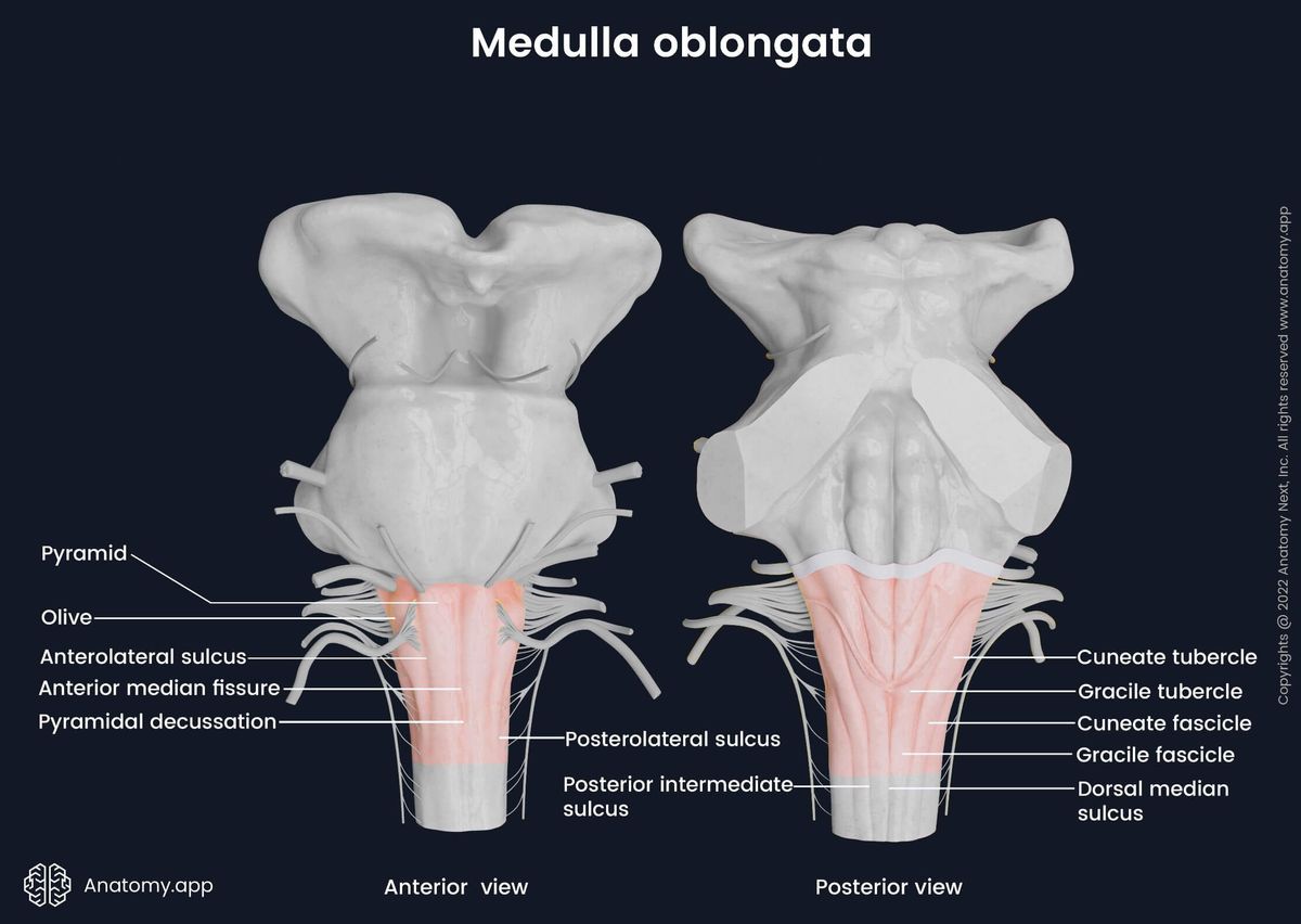
The most noticeable features of the ventral surface are two paired prominences - pyramids (2) and olives (2) - and five horizontal grooves. The five grooves (one fissure and four sulci) separate the previously mentioned prominences, and they include the following:
- Anterior median fissure
- Left and right anterolateral sulci
- Left and right posterolateral sulci
The anterior median fissure runs along the midline of the ventral surface, and it separates both pyramids. The anterior median fissure is a continuation of the anterior median fissure of the spinal cord. On each lateral side of the pyramids extend the anterolateral sulcus. This groove separates the pyramid from the olive on each side. Laterally to each olive, additional groove called the posterolateral sulcus runs parallel to the previously mentioned sulcus.
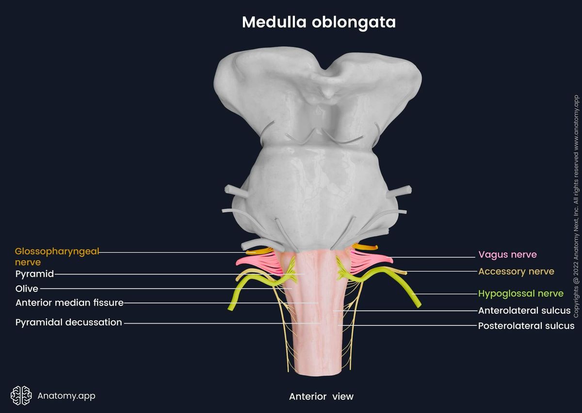
The pyramids are paired prominences located between the anterior median fissure and the anterolateral sulci. The pyramids are primarily formed by the fibers of the white matter as the corticospinal tracts pass through them. Near the junction of the medulla oblongata and the spinal cord, the motor fibers of the corticospinal tracts cross in the midline, forming the pyramidal decussation.
Next to the pyramids, on the ventral surface of the medulla oblongata are situated two subsequent eminences called the olives. They are located between the anterolateral and posterolateral sulci. Internally they house the inferior olivary nuclei. Moreover, the hypoglossal nerve (CN XII) exits on the anterolateral sulcus between the olive and pyramid.
Overall, four paired cranial nerves emerge on the ventral surface of the medulla oblongata. As previously mentioned, the hypoglossal nerves (CN XII) arise on the anterolateral sulci. The glossopharyngeal (CN IX), vagus (CN X), and accessory (CN XI) nerves exit the brain via the most lateral located grooves of the medulla oblongata - the right and left posterolateral sulci.
Dorsal surface of medulla oblongata
The dorsal surface of the medulla oblongata is the posterior external surface that faces the cerebellum. It is connected to the cerebellum by the inferior cerebellar peduncles. The dorsal surface forms the inferior part of the rhomboid fossa and faces the fourth ventricle.
The obex is the most caudal part of the fourth ventricle, where it narrows and transitions into the central canal of the spinal cord. Thus, the central canal of the spinal cord is a direct continuation of the fourth ventricle. Inferiorly, between the dorsal surface of the medulla oblongata and the anterior aspect of the cerebellum is the cisterna magna, also called the cerebellomedullary cistern.
The cisterna magna is the largest of all subarachnoid cisterns. It is an unpaired expansion of the subarachnoid space filled with cerebrospinal fluid (CSF). The cisterna magna connects to the fourth ventricle via the median (foramen of Magendie) and lateral (foramina of Luschka) apertures. It houses the vertebral arteries, glossopharyngeal nerve (CN IX), vagus nerve (CN X), accessory nerve (CN XI) and the CSF producing choroid plexus.
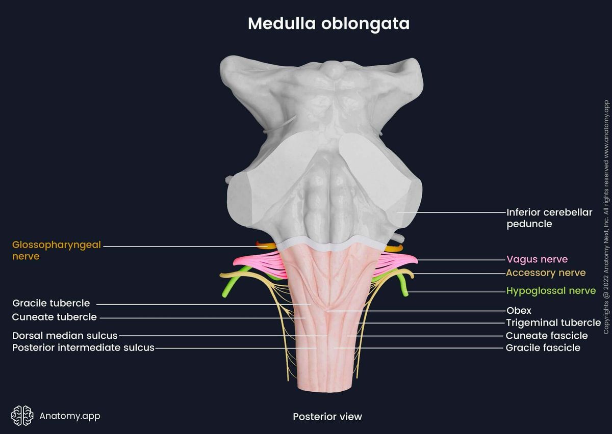
A shallow groove goes along the midline of the dorsal surface, which is called the dorsal median sulcus. The dorsal median sulcus, also known as the posterior median sulcus, is a continuation of the posterior median sulcus of the spinal cord. The posterior median sulcus ends approximately in the middle of the medulla oblongata, where the central canal widens into the fourth ventricle.
Lateral on both sides of the dorsal median sulcus are paired eminences known as fascicles. On both sides of the groove is situated the gracile fascicle, while lateral to it is the cuneate fascicle. Superiorly, the fascicles form two prominences: the gracile and cuneate tubercles. The gracile tubercle houses a part of the gracile nucleus, and the cuneate tubercle contains the upper part of the cuneate nucleus. Lateral to the cuneate tubercle is another prominence called the trigeminal tubercle. Beneath it is the spinal nucleus of the trigeminal nerve (CN V).
The dorsal surface of the medulla oblongata contains two more grooves - right and left posterior intermediate sulci. They are situated on both sides of the posterior median sulcus parallel to it. Inferiorly, the posterior intermediate sulcus separates the cuneate and gracile fascicles. In the superior direction, the margins of the dorsal surface become thicker and form the inferior cerebellar peduncles.
Medulla oblongata internal anatomy
The internal anatomy of the medulla oblongata is very complex as it contains a multitude of nuclei (clusters of neuron cell bodies) and tracts (consisting of nerve fibers). When looked at in cross-section, the medulla oblongata comprises three separate parts: basis of medulla oblongata, tegmentum, and tectum.
The basis is the most ventral part of the medulla oblongata, and it contains the pyramids with the pyramidal decussation. The tegmentum is the middle portion, and it is continuous with the tegmentum of the pons. The tegmentum contains most of the tracts and nuclei (inferior olivary nuclei, nuclei of the cranial nerves IX, X, XI, XII). The tectum is the most dorsal part of the medulla. It is composed of the inferior medullary velum - the most inferior and posterior aspect of the fourth ventricle.
As with any other part of the brain, the medulla oblongata consists of grey and white matter. The grey matter is composed of neuronal cell bodies that form nuclei. The white matter is comprised of neuronal axons (creating nerve fibers), which are grouped into ascending and descending collections of fibers known as the tracts. The neural tracts and axons connect the nuclei of the medulla oblongata with different regions and structures of the central nervous system (CNS).
Basis of medulla oblongata
The basis is the most ventral part of the medulla oblongata, and it contains the pyramids. The pyramids are white matter structures composed of the corticospinal and corticobulbar tract fibers. Both pathways together create the so-called pyramidal tract. Below the pyramids in the anterior median fissure region, the majority of the corticospinal tract fibers cross, forming the pyramidal decussation. Besides the structures of the white matter, the basis of the medulla oblongata also contains nuclei of the gray matter. On the anterior surface of each pyramid, a collection of neurons called the arcuate nucleus is located.
Pyramidal tract
The pyramidal tract consists of the corticospinal and corticobulbar fibers. It is a descending pathway responsible for voluntary movements of the limbs and trunk (corticospinal fibers) and face, head and neck (corticobulbar fibers). The corticospinal fibers originate from the cortex and regulate voluntary movements through the neurons of the spinal cord. Approximately 80-90% of the fibers cross and form the pyramidal decussation.
The pyramidal decussation is the major decussation site of the descending motor corticospinal fibers. It is located in the anterior lower part of the basis of the medulla oblongata. After crossing, the fibers form the lateral corticospinal tract that further descends to reach the spinal cord. Fibers that do not cross in the midline become the anterior corticospinal tract.
The corticobulbar fibers also called the corticonuclear fibers, regulate voluntary movements with the help of the cranial nerves. Within the medulla oblongata, they synapse with the motor nuclei of the glossopharyngeal (CN IX), vagus (CN X), accessory (CN XI), and hypoglossal (CN XII) cranial nerves.
Arcuate nucleus
The arcuate nucleus is a group of smaller nuclei. It is a bilaterally located relay nucleus situated on the anterior surface of each pyramid. The arcuate nucleus receives information from the corticospinal tract and passes it to the cerebellum with the help of the anterior external arcuate fibers and striae medullares via the inferior cerebellar peduncle.
Tegmentum of medulla oblongata
The tegmentum of the medulla oblongata is a direct extension of the tegmentum of the pons and midbrain. It is the most significant part of the medulla oblongata, housing the majority of the nuclei and descending and ascending tracts. The tegmentum of the medulla oblongata contains three groups of nuclei:
- Cranial nerve nuclei
- Relay nuclei
- Reticular nuclei
Cranial nerve nuclei
The cranial nerve nuclei are paired sensory or motor nuclei of the cranial nerves. They form afferent and efferent connections with the respective anatomical structures including higher brain regions, often through the thalamus. Located in the medulla oblongata are nuclei of the trigeminal (CN V), facial (CN VII), vestibulocochlear (CN VIII), glossopharyngeal (CN IX), vagus (CN X), accessory (CN XI), and hypoglossal (CN XII) nerves. And these nuclei include the following:
- Spinal nucleus of the trigeminal nerve
- Vestibular nuclei (medial, lateral, inferior)
- Nucleus ambiguus
- Nucleus of the solitary tract
- Inferior salivatory nucleus
- Dorsal nucleus of the vagus nerve
- Nucleus of the hypoglossal nerve
The spinal nucleus of the trigeminal nerve is a general somatic sensory nucleus situated in the posterolateral tegmentum. It descends into the medulla oblongata from the caudal end of the principal sensory nucleus of the trigeminal nerve located in the pons. The spinal nucleus receives afferent nerve fibers. They carry information about touch, vibration, pain, and temperature from the ipsilateral side of the face. The spinal nucleus of the trigeminal nerve is composed of three subdivisions - the oral, caudal, and interpolar parts (subnuclei).
The vestibular nuclei of the vestibulocochlear nerve (CN VIII) are situated in both - pons and medulla oblongata. There are four paired vestibular nuclei: superior vestibular nucleus, medial vestibular nucleus, lateral vestibular nucleus, and inferior vestibular nucleus. The superior vestibular nucleus is located in the pons, while the other three are positioned in the tegmentum of the medulla oblongata.
The medial vestibular nucleus is located in the vestibular area of the medulla oblongata close to the surface of the fourth ventricle’s floor. The lateral vestibular nucleus is situated lateral to the previous nucleus in the upper medulla, while the inferior vestibular nucleus is positioned in the lower part of the medulla oblongata inferior to the lateral vestibular nucleus. The vestibular nuclei receive sensory input from the vestibular portion of the vestibulocochlear nerve (CN VIII) and regulate head position, motion, spatial orientation, equilibrium, and posture.
The nucleus ambiguus is a special visceral motor nucleus of the glossopharyngeal (CN IX), vagus (CN X), and accessory (CN XI) nerves. It is a motor nucleus situated deep within the reticular formation. Efferent fibers from the nucleus ambiguus innervate the muscles of the soft palate, pharynx, larynx, and upper esophagus. Hence, it partakes in regulating speaking and swallowing. Moreover, the nucleus ambiguus is part of the parasympathetic system and modulates heart rate.
The nucleus of the solitary tract is a general and special visceral sensory nucleus of the facial (CN VII), glossopharyngeal (CN IX), and vagus (CN X) nerves. It is located in the posterior part of the tegmentum. It receives general visceral information from the mechanoreceptors and chemoreceptors in the heart, lungs and airways, gastrointestinal tract, pharynx, liver, nasal cavity, paranasal sinuses, and soft palate.
The inferior salivatory nucleus of the glossopharyngeal nerve (CN IX) is a general visceral motor (parasympathetic) nucleus. It is situated in the anterior part of the tegmentum below the pontomedullary junction and inferior to the superior salivatory nucleus. The parasympathetic fibers arising from the inferior salivatory nucleus travel within the glossopharyngeal nerve and innervate the parotid gland through the otic ganglion.
The dorsal nucleus of the vagus nerve is a general visceral motor (parasympathetic) nucleus located in the posterior aspect of the tegmentum. The fibers originating from the nucleus innervate and regulate the functioning of the heart, lungs, bronchi, esophagus, stomach, small intestine, cecum, ascending colon and two-thirds of the transverse colon.
The nucleus of the hypoglossal nerve is a prominent elongated column of motor nuclei. It is situated in the posterior part of the tegmentum of the medulla oblongata medial to the dorsal nucleus of the vagus nerve. From the nucleus of the hypoglossal nerve, efferent nerve fibers arise that innervate all intrinsic and extrinsic tongue muscles, except for the palatoglossus muscle. The latter is innervated by the vagus nerve (CN X).
Relay nuclei
The paired relay nuclei are bilaterally scattered throughout the tegmentum of the medulla oblongata. They form extensive synaptic networks. Information in the form of stimuli is further sent from the peripheral receptor areas to the higher cortical centers. The relay nuclei located in the tegmentum include:
- Cuneate nucleus
- Gracile nucleus
- Inferior olivary nucleus
The cuneate nucleus is located in the dorsolateral portion of the tegmentum. It is a sensory nucleus partly found within the cuneate tubercle. Via the inferiorly situated cuneate fasciculus, the cuneate nucleus receives sensory information about touch, conscious proprioception, and vibration from the upper limb and upper trunk. The fibers that originate from the cuneate nucleus decussate and join the medial lemniscus.
The gracile nucleus is situated medially to the cuneate nucleus. It is a sensory nucleus partly located within the gracile tubercle. Via the gracile fasciculus, the gracile nucleus receives afferent information about fine touch, conscious proprioception, and vibration from the lower limb and lower trunk. The fibers from the nucleus decussate and join the medial lemniscus.
The inferior olivary nucleus is a prominent nucleus located posterior to each pyramid. It is composed of three smaller nuclei: dorsal and medial accessory olivary nuclei and the principal inferior olivary nucleus. The inferior olivary nucleus coordinates signals transmitted from the spinal cord to the cerebellum. Therefore, it regulates motor coordination and learning.
Reticular nuclei
The reticular nuclei are a part of the reticular formation. The reticular nuclei of the medulla oblongata are located within the medullary reticular formation, which is found centrally within the tegmentum. The reticular formation is an interconnected network of brainstem nuclei and neurons. It is the site of integration and relay of vital information from the cerebrum, cerebellum and spinal cord. The reticular nuclei of the medulla oblongata include:
- Raphe nuclei
- Gigantocellular nuclei
- Perihypoglossal nuclei
- Lateral reticular nucleus
The raphe nuclei are located near the midline throughout the entire length of the brainstem and are considered to be the primary source of the neurotransmitter serotonin. The raphe nuclei participate in the modulation of mood, pain, arousal state, and thermoregulation.
The gigantocellular nucleus is situated in the ventromedial part and gives fibers that connect with the hypoglossal nucleus. The perihypoglossal nuclei take part in modulating eye movements. They receive information from the cerebral cortex, vestibular nuclei, accessory oculomotor nuclei and transfer it to the cerebellum, thalamus, and cranial nerve nuclei.
The lateral reticular nucleus is located bilaterally posterolateral to the inferior olivary nucleus, and it receives information from a wide array of sources. Subsequently, it sends information to the ipsilateral hemisphere of the cerebellum and participates in the regulation of motor functions.
Ascending tracts of tegmentum
The white matter of the tegmentum forms ascending and descending tracts. Most of the following pathways only pass through the medulla oblongata, but some of them also originate from it. The ascending tracts of the tegmentum include:
- Gracile fasciculus
- Cuneate fasciculus
- Spinothalamic fibers
- Posterior spinocerebellar tract
- Anterior spinocerebellar tract
- Olivocerebellar tract
- Spinal tract of the trigeminal nerve
The gracile fasciculus is located inferior to the gracile tubercle and nucleus. It conveys fine tactile, vibratory, and conscious proprioceptive information. Signals are received from the lower limbs and lower parts of the trunk. The cuneate fasciculus receives fine tactile, vibratory, and conscious proprioceptive information from the upper limbs and upper trunk.
The cuneate and gracile fasciculi contain nerve fibers from the sensory dorsal root ganglia of the spinal nerves (first-order neurons). These nerve fibers ascend and reach cuneate and gracile nuclei in the medulla oblongata (second-order neurons). The axons of these nuclei form the internal arcuate fibers that cross and form the so-called sensory decussation (decussation of the medial lemniscus). After the decussation, the fibers form the medial lemniscus and via the thalamus (third-order neurons) send information about conscious proprioception and fine touch to the primary somatosensory cortex.
The spinothalamic fibers pass through the anterolateral medulla. They are formed by the lateral and anterior spinothalamic tracts. The fibers transmit sensory information about pain, pressure, temperature, and crude touch to the thalamus. The anterior spinothalamic tract connects the nucleus proprius and substantia gelatinosa in the spinal cord with the thalamus. It carries information about crude touch and pressure sensation.
The lateral spinothalamic tract is an ascending pathway that connects the substantia gelatinosa and nucleus proprius in the spinal cord with the thalamus. From the thalamus, the information is passed to the cerebral cortex (primary somatosensory cortex). The lateral spinothalamic tract carries pain and temperature sensations.
The posterior spinocerebellar tract is situated in the posterolateral part of the tegmentum. It conveys information about unconscious proprioception, touch, and pressure from the lower limbs and trunk to the cerebellum via the inferior cerebellar peduncles.
The anterior spinocerebellar tract is located in the anterolateral part of the tegmentum anterior to the posterior spinocerebellar tract. It transmits unconscious proprioceptive information from the lower limbs to the cerebellum via the superior cerebellar peduncles.
The olivocerebellar tract is a pathway that passes in the upper anterior part of the tegmentum, transmitting information about motor coordination from the inferior olivary nucleus to the cerebellum.
The spinal tract of the trigeminal nerve is located in the posterolateral tegmentum of the medulla oblongata. Medial to this tract is the spinal nucleus of the trigeminal nerve. This pathway carries the sensory information from the face to the spinal nucleus of the trigeminal nerve.
Descending tracts of tegmentum
Besides the ascending tracts, several descending pathways pass through the tegmentum of the medulla oblongata. The descending tracts travel from the higher regions of the brain to the lower parts of the spinal cord. The descending tracts of the tegmentum include the following:
- Rubrospinal tract
- Tectospinal tract
- Reticulospinal tract
- Medial longitudinal fasciculus
- Vestibulospinal tract
- Olivospinal tract
The rubrospinal tract originates from the red nucleus in the midbrain and descends through the pons and medulla oblongata to reach the motor nuclei of the spinal cord. It is a part of the extrapyramidal system. In the medulla oblongata, this pathway is bilaterally situated on both lateral sides of the tegmentum. This tract regulates and coordinates balance automatic movements and controls the muscle tone of the upper limb flexors.
The tectospinal tract is a motor pathway and part of the extrapyramidal system. It originates from the superior colliculus of the midbrain and descends to the motor nuclei of the upper cervical spinal cord levels. The tectospinal tract is located anterior to the medial longitudinal fasciculus and posterior to the medial lemniscus. It transmits impulses that coordinate reflector head and eye movements as a protective reaction to unexpected visual or auditory stimuli.
The reticulospinal tract is composed of two pathways - the pontine (medial) and medullary (lateral) reticulospinal tracts. This tract is a part of the extrapyramidal system. It helps in maintaining posture and balance, and this tract also coordinates stimulation and inhibition of extensors and flexors of the limbs. The reticulospinal tract connects the reticular formations located within the brainstem with the reticular formations and motor nuclei of the spinal cord.
The medial longitudinal fasciculus is a sizable nerve bundle situated in the central part of the posterior tegmentum. It connects the motor nuclei of the cranial nerves CN III (oculomotor), CN IV (trochlear), CN VI (abducens), and CN VIII (vestibulocochlear) with each other and with the motor nuclei of the spinal cord’s cervical and upper thoracic segments. Together with the medial vestibulospinal and tectospinal tracts, the medial longitudinal fasciculus coordinates eye movements and associated head and neck movements.
The vestibulospinal tract is a part of the extrapyramidal system, and it is made up of the lateral and medial vestibulospinal tracts. It maintains reflexes linked to the balance with the help of the vestibular input. The vestibulospinal tract connects the vestibular nuclei of the pons and medulla oblongata with the motor nuclei of the spinal cord.
The olivospinal tract is also a part of the extrapyramidal system. It is a descending pathway connecting the inferior olivary nuclei of the medulla oblongata with the cervical segments of the spinal cord. This tract provides reflex movements arising from proprioception. However, its existence is doubtful. Some authors claim that the olivospinal tract is a part of the spino-olivary pathway because it contains fibers of the olivospinal tract.
Cross-section of medulla oblongata
To better understand the structures of the medulla oblongata and their relations, the medulla oblongata is typically examined in three cross-section levels. From the most inferior to the most superior, these levels are: pyramidal decussation level, sensory decussation level and the level of the olives.
Some structures are seen in all cross-sections, and others only in one. Apart from a few minor changes in the location, many structures, especially tracts, are present at all three levels. Nuclei, as they tend to be smaller in size, are often only seen in one of the cross-sections.
For instance, the olivocerebellar tract is only visible at the topmost cross-sections - at the level of the olives. Moreover, the gracile and cuneate fasciculi are present in the two lower cross-sections, respectively, at the level of the sensory decussation and pyramidal decussation.
Whereas the corticospinal, anterior and lateral spinothalamic, posterior and anterior spinocerebellar, rubrospinal, tectospinal reticulospinal and vestibulospinal tracts, as well as the medial longitudinal fasciculus, are seen at all levels, as they pass through the entire length of the medulla oblongata.
Level of pyramidal decussation
At the caudal part of the medulla oblongata, the corticospinal tract fibers that are found within the pyramids cross over (decussate), and this is known as the decussation of the pyramids or pyramidal decussation. The lower medulla oblongata is the point of reorganization of spinal cord structures to those of the brainstem. Moreover, the central canal of the spinal cord can be seen in the center of the lower medulla oblongata.
The pyramidal decussation is a significant decussation site of the descending motor corticospinal fibers. It is located in the lower anterior aspect of the basis, right posterior to the anterior median fissure. Approximately 80-90% of the corticospinal fibers cross and form the pyramidal decussation.
As corticospinal fibers enter the decussation, the pyramids progressively reduce in volume in the downward direction. After crossing, the fibers form the lateral corticospinal tract that further descends to reach the spinal cord. The 10-20% of fibers that do not cross in the midline become the anterior corticospinal tract.
At the level of the pyramidal decussation, the gracile and cuneate nuclei are found within the dorsal tegmentum of the medulla oblongata. Both nuclei are located anterior to their respective fasciculi and posterior median sulcus. The cuneate and gracile fasciculi ascend from the dorsal column of the spinal cord.
Posterolaterally in the tegmentum of the medulla oblongata is the spinal nucleus of the trigeminal nerve and its tract. The spinal tract of the trigeminal nerve is situated lateral to the spinal nucleus. Additionally, the medial longitudinal fasciculus is located on both lateral sides of the pyramidal decussation near the spinothalamic tracts.
Level of sensory decussation
At the level of the sensory decussation (decussation of the medial lemniscus), the gracile and cuneate fasciculi continue to ascend within the dorsal tegmentum. The internal arcuate fibers originate from the gracile and cuneate nuclei. These fibers leave the respective nuclei and go in the anterior direction.
Lateral to the internal arcuate fibers is the central canal, hypoglossal nucleus, and the medial longitudinal fasciculus (MLF). When passing along the MLF, the internal arcuate fibers turn in the medial direction and decussate. The decussation of the internal arcuate fibers is also known as sensory decussation. After the fibers cross, they form the medial lemniscus that ascends to the thalamus.
The sensory decussation is located dorsal to the pyramids and ventral to the central grey matter. Also, the pyramidal tract is located anterior to the decussation, while the medial longitudinal fasciculus is situated posterior to it. Moreover, at this level, the hypoglossal nuclei are situated in the posterior aspect of the tegmentum, close to the midline.
Anterior to the gracile nucleus is the dorsal motor nucleus of the vagus nerve, the solitary tract, and the nucleus of the solitary tract. Lateral to the medial lemniscus and posterior to the inferior olivary nucleus is the nucleus ambiguus. The lower part of the inferior olivary nucleus is situated lateral to the medial lemniscus, posterior to the pyramids and anterior to the nucleus ambiguus.
Level of olives
At the level of the olives, the medulla oblongata is significantly wider. Centrally along the midline, three pathways - the medial lemniscus, medial longitudinal fasciculus and tectospinal tract - are found. The central canal has moved more posteriorly and transformed into the fourth ventricle. Therefore, at this level, the medulla oblongata has become the open medulla.
In the anterior part of the medulla oblongata, the pyramids and prominent olives can be seen. The arcuate nucleus is found within the anterior aspect of the pyramid. Within the olives, the large inferior olivary nucleus is located. The anterior spinocerebellar tract is situated in front of the inferior cerebellar peduncles. Slightly to the sides, the anterior (ventral) cochlear nucleus is located in the anterior aspect of the inferior cerebellar peduncle.
Posterolaterally, the lower parts of the inferior cerebellar peduncles can be seen. Around their base, many nuclei are located. The spinal nucleus of the trigeminal nerve and its tract is located medially to the peduncle. Posteriorly to the peduncle is the inferior vestibular nucleus, and behind it are the smaller posterior (dorsal) cochlear and medial vestibular nuclei.
In the posterior part of the tegmentum, anterior to the fourth ventricle, the medial longitudinal fasciculus and the hypoglossal nucleus have migrated backward. The dorsal motor nucleus of the vagus nerve and the nucleus of the solitary tract are located lateral to the hypoglossal nucleus.
Vasculature of medulla oblongata
The medulla oblongata is supplied by the anterior and posterior spinal arteries, anterior and posterior inferior cerebellar arteries, and the vertebral artery. The anterior and posterior spinal arteries and posterior inferior cerebellar artery are branches of the vertebral artery. In contrast, the anterior inferior cerebellar artery is a branch of the basilar artery. The venous drainage is provided by the ventral, ventrolateral, lateral, and posterior medullary veins.
The anterior spinal artery supplies the lower part of the anterior medulla oblongata and the ventral border of the fourth ventricle. Moreover, its medullary arteries are also widely distributed within the medulla oblongata. The posterior spinal artery supplies the rear part of the medulla oblongata below the olives.
The posterior inferior cerebellar artery provides arterial blood supply to the dorsal medulla oblongata at the level of the olives. The remaining parts of the medulla oblongata are supplied directly by the vertebral arteries and anterior inferior cerebellar arteries.
The ventral (anterior), ventrolateral (anterolateral), lateral and posterior medullary veins ensure venous drainage from the medulla oblongata. These veins further drain into the occipital sinus dorsally and into the basilar venous plexus and inferior petrosal sinus ventrally.
References:
- Byrne, J. V. (2017). Tutorials in Endovascular Neurosurgery and Interventional Neuroradiology (2nd ed.). Springer.
- Crossman, A. R., & Neary, D. (2019). Neuroanatomy: an Illustrated Colour Text (6th ed.). Elsevier.
- Goetz, C. (2007). Textbook of Clinical Neurology (3rd edition). Saunders.
- Iordanova, R., & Reddivari, A. K. R. (2021). Neuroanatomy, Medulla Oblongata. NCBI. https://www.ncbi.nlm.nih.gov/books/NBK551589/
- Standring, S. (2020a). Brainstem. In Gray’s Anatomy: The Anatomical Basis of Clinical Practice (42nd ed., pp. 442–464). Elsevier.
- Standring, S. (2020b). The anatomy of the vascular and lymphatic systems. In Gray’s Anatomy: The Anatomical Basis of Clinical Practice (42nd ed., p. 1464). Elsevier.
- Vanderah, T. W., & Gould, D. J. (2020). Nolte’s The Human Brain (8th ed.). Elsevier.
Anatomy.app
Contact information
- For questions regarding business inquiries. Please contact:
- info@anatomy.app