- Anatomical terminology
- Skeletal system
- Joints
- Muscles
- Heart
- Blood vessels
- Nervous system
- Respiratory system
- Digestive system
- Lymphatic system
- Female reproductive system
- Male reproductive system
- Endocrine glands
- Eye
- Ear
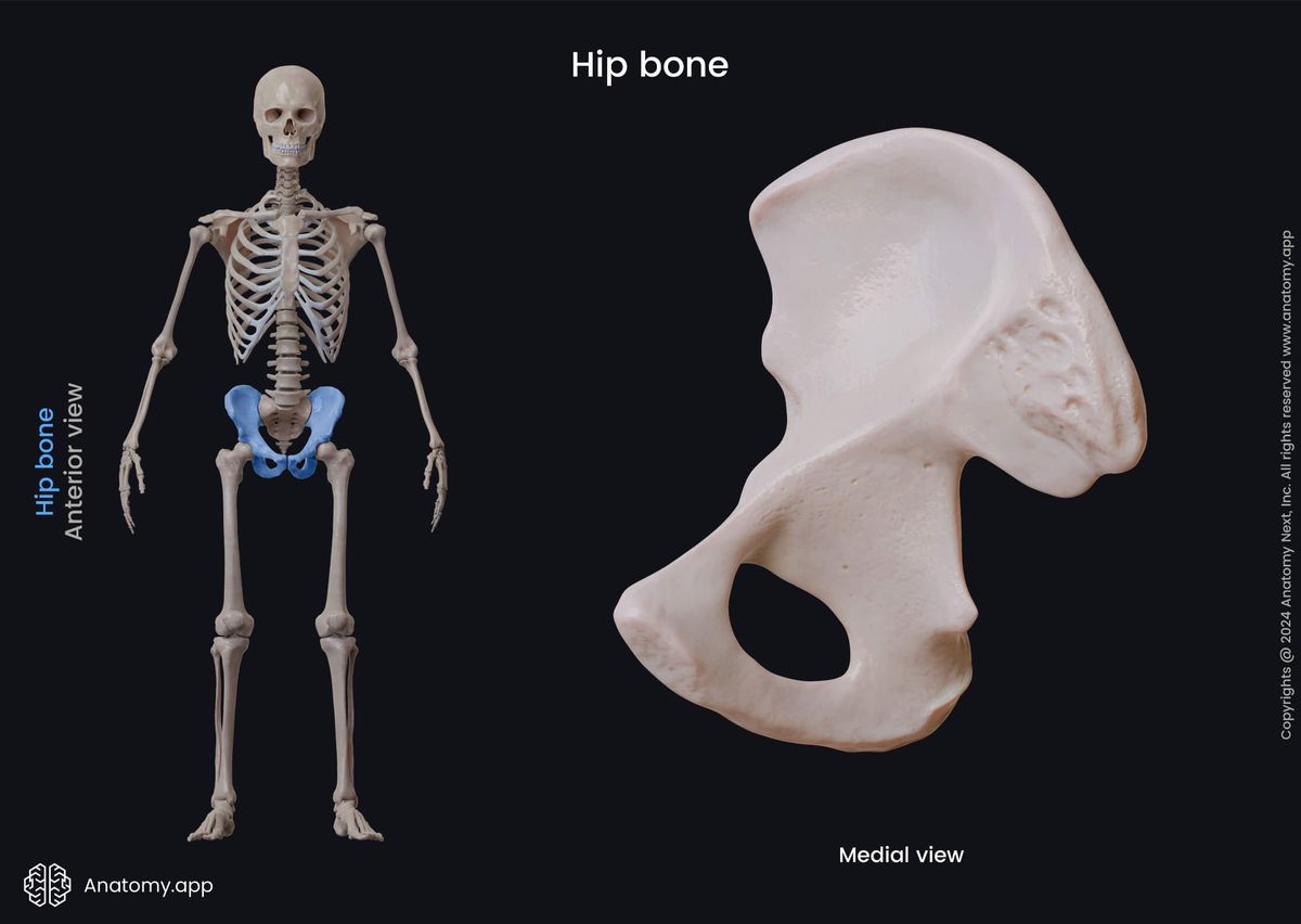
Hip bone
The hip bone (Latin: os coxae), also known as the pelvic or coxal bone, is a paired anatomical structure formed by the fusion of three bones - the ilium, ischium, and pubis. The left and right hip bones join together at the pubic symphysis.
While a person is growing, all three mentioned bones that form the hip bone are separated by cartilages that gradually ossify. All three bones usually fuse around the age of 15 to 17 and during puberty, but they ossify around 25 years.
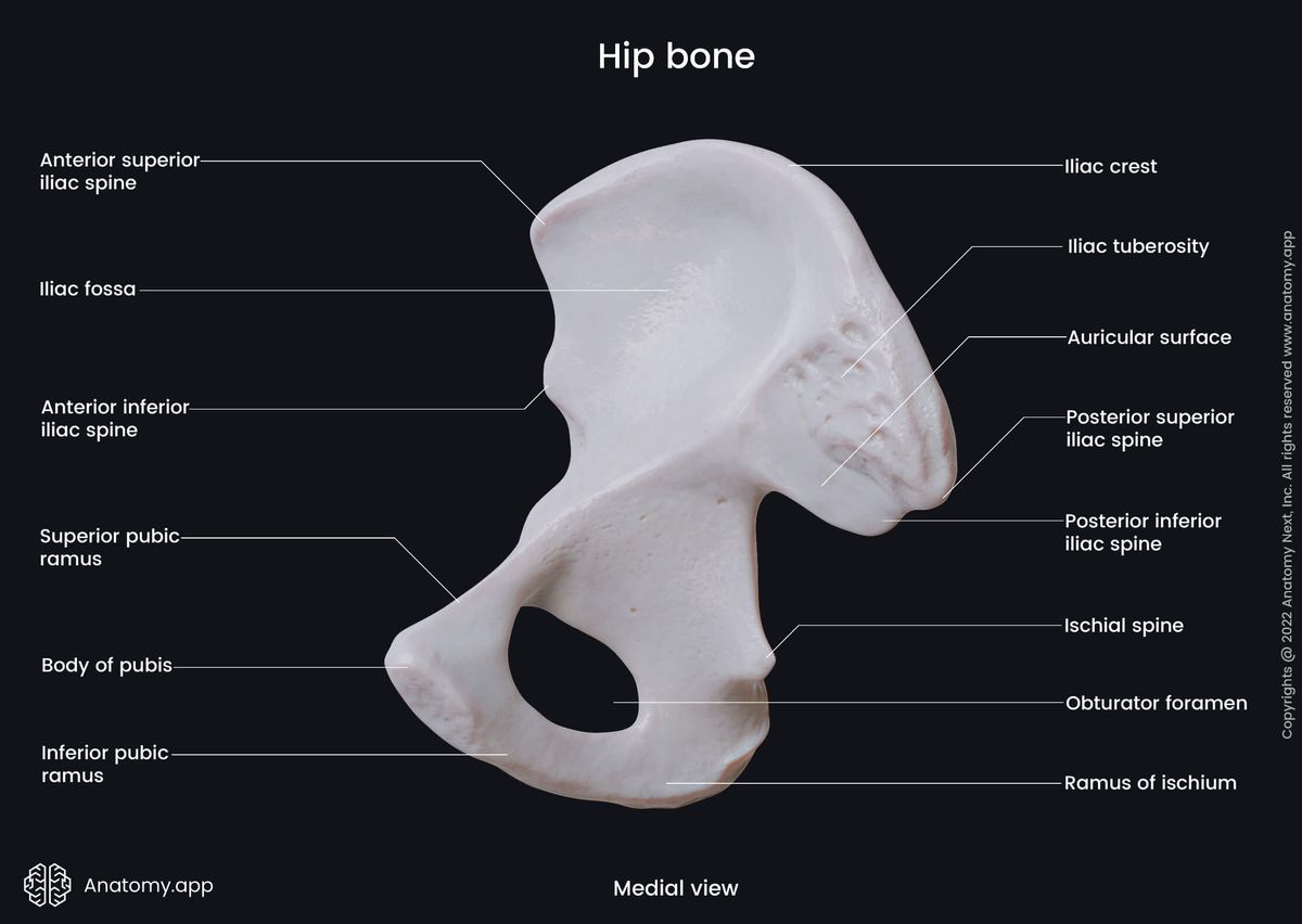
The ilium or iliac bone is a paired bone that forms the uppermost and most significant part of the hip bone. It consists of a body, which is the central part, and a wing - an extended superior portion of the bone.
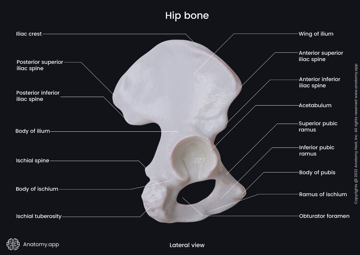
The ischium is a paired bone forming the lower and posterior part of the hip bone. It also forms the posterior and inferior aspects of the obturator foramen. The ischium presents two main parts - a body and a ramus.
The pubis or pubic bone is a paired bone forming the anterior part of the hip bone. It is composed of three main parts - a body and two rami (superior and inferior rami). The left and right pubic bones join at the pubic symphysis.
The fusion of all three parts of the hip bone creates several anatomical landmarks located between them. These landmarks include the acetabulum located on the external surface of the hip bone and the obturator foramen.
Acetabulum
The acetabulum is a deep, cup-shaped cavity located on the external surface of the hip bone. It is the site where fits the head of the femur, forming the hip joint. The acetabulum is also the site where all three bones that form the hip bone fuse.
The ischium forms the lower posterior aspect (one-third) of the acetabulum, while the ilium forms the superior one-third of it. The anterior one-third of the cavity is formed by the pubis. The acetabulum features the following landmarks:
- Lunate surface
- Acetabular fossa
- Acetabular notch
- Iliopubic eminence
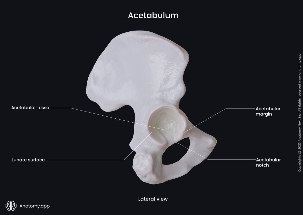
The lunate surface of the acetabulum is a curved articular surface surrounding the acetabular fossa. It is located above and on the sides of the fossa. The lunate surface articulates with the head of the femur.
The acetabular fossa is a non-articular depression located at the center of the acetabulum. The fossa is occupied by the ligament of the head of femur. Inferiorly, the acetabular fossa is continuous with the acetabular notch.
The acetabular notch is a deep indentation between both ends of the lunate surface below the acetabular fossa. The edges of this notch serve as attachment sites for the ligament of the head of the femur. The transverse acetabular ligament stretches above the notch and transforms it into a foramen, through which nerves and blood vessels reach the hip joint.
The iliopubic eminence or iliopectineal eminence is a flat prominence at the proximal part of the pubis above the acetabulum where the pubic and iliac bones fuse. It is also the medial border of the groove, over which the iliacus and psoas major muscles pass.
Obturator foramen
The obturator foramen is a large opening on each side of the pelvis formed by the fusion of the ischium and pubis. It serves as the passage site for the obturator artery, vein and nerve exiting the pelvis.
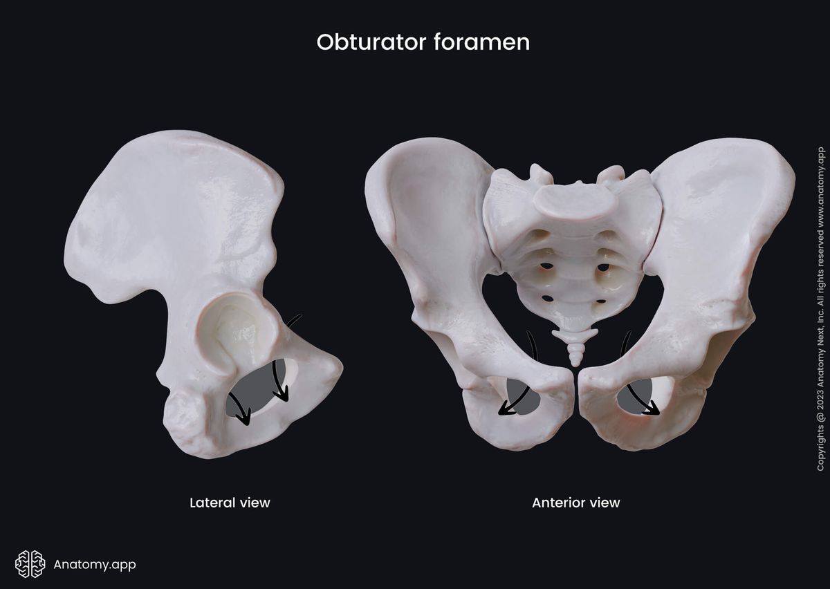
This opening has a thin, uneven margin, which presents with a deep obliquely groove called the obturator groove. This groove runs on the inferior aspect of the superior ramus of pubis in a medial and inferior direction.
The obturator groove is transformed into the obturator canal with the help of a ligamentous band that is an upper part of the obturator membrane. This ligamentous band attaches to two tubercles:
- Posterior obturator tubercle - a bony eminence located at the end of the obturator groove below it;
- Anterior obturator tubercle - a bony eminence found at the end of the obturator groove above and anteriorly to it.
Anatomy.app
Contact information
- For questions regarding business inquiries. Please contact:
- info@anatomy.app