- Anatomical terminology
- Skeletal system
- Joints
- Muscles
- Heart
- Blood vessels
- Nervous system
- Respiratory system
- Digestive system
- Lymphatic system
- Female reproductive system
- Male reproductive system
- Endocrine glands
- Eye
- Ear
Heart
The heart (Latin: cor) is a thick, muscular organ with four cavitated parts located in the middle mediastinum. It is a central organ and pump of the cardiovascular system that maintains the unidirectional flow of blood through the circulatory system. The heart is a vital organ meaning a person can not live without it.
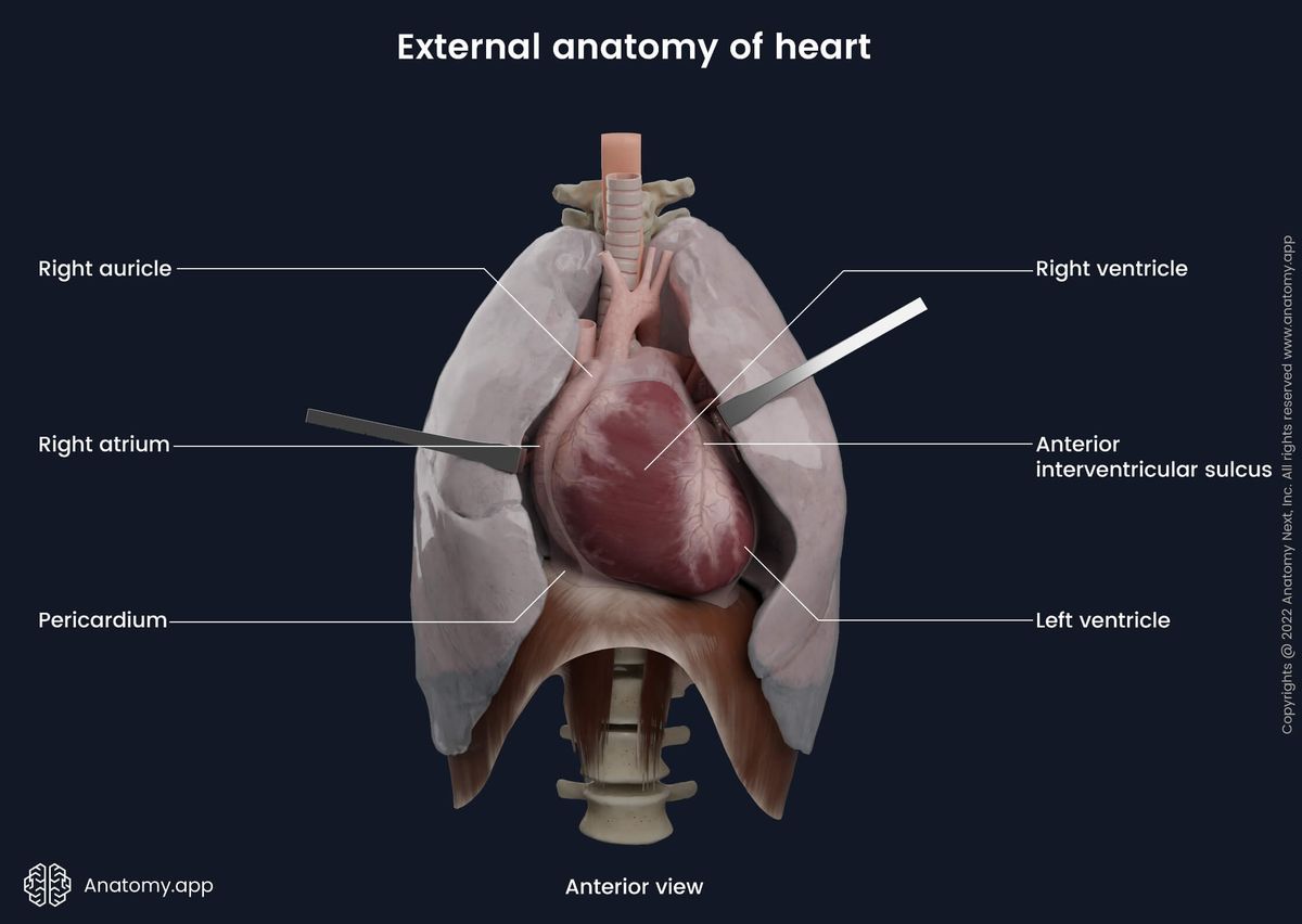
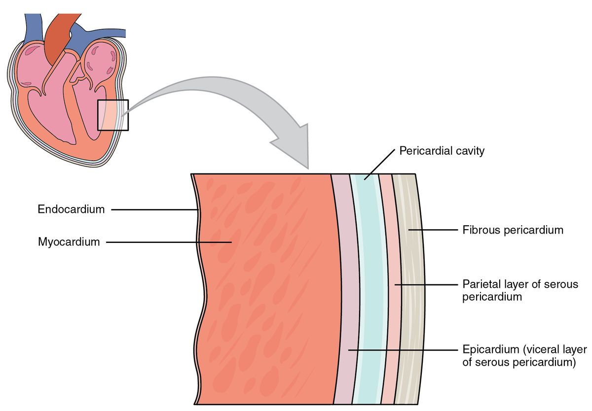
The heart consists of the right and left side (or right and left pump) and four main parts: right atrium and right ventricle, left atrium and left ventricle. The heart is surrounded by a serous sac called the pericardium. The great vessels originating from the heart provide blood flow throughout the whole body.
Heart sound locations and heart auscultation
The closing of each heart valve can be auscultated at a specific place on the anterior wall of the chest.
- The right atrioventricular or tricuspid valve can be auscultated in the fifth intercostal space on the right side of the sternum or the xiphoid process of the sternum.
- The sound of the left atrioventricular or mitral valve can also be best heard in the fifth intercostal space on the left side, approximately 1,5 centimeters medial from the left midclavicular line.
- The aortic valve can be found in the second intercostal space on the right side of the sternum.
- The pulmonary valve can also be heard in the second intercostal space but on the left side of the sternum.
Pulmonary and systemic circulation
The systemic circulation starts in the left ventricle containing oxygenated blood (rich in oxygen). Through the aorta and its branches, blood spreads through the body and reaches all organs. Tissue receives oxygen, and the oxygenated blood becomes deoxygenated. It is being accomplished by the arterioles, pre-capillaries, capillaries, post-capillaries, and finally by the venules going back to the heart. Deoxygenated blood reaches the heart through the superior and inferior venae cavae and coronary sinus, flowing into the right atrium, where ends the systemic circulation.
The pulmonary circulation starts with the right ventricle containing deoxygenated blood. Blood reaches the lungs through the pulmonary trunk and right and left pulmonary arteries. Arteries form a network of capillaries in the lungs and in the alveolar walls where happens the gas exchange. Deoxygenated blood becomes oxygenated and reaches the left atrium of the heart through the pulmonary veins. The pulmonary circulation ends in the left atria of the heart.
Heart functions
The heart works as a pump of the circulation system. Its main function is to pump the blood to the systemic and pulmonary circulations providing exchange of deoxygenated and oxygenated blood. Therefore, the heart provides the tissue with oxygen. The heart is the main organ in the circulatory system, and its rhythmic contractions keep an individual alive.
- The heart generates blood pressure.
- The heart routes the blood as it separates the pulmonary and systemic circulations.
- It ensures one-way blood flow because of the valves.
- The heart regulates the blood supply as it changes contraction rate and force, responding to metabolic needs.
- It provides the cardiac cycle.
Cardiac cycle
The rhythmic action of the heart and, therefore, the blood flow to the systemic and pulmonary circulations are provided by the sequential contractions of the atria and ventricles. This is called the cardiac cycle. The cycle starts with one heartbeat and ends with another.
Each cycle can be divided into two main phases - diastolic and systolic phases. During the diastolic phase, a heart chamber is relaxed, and it fills with blood. During the systolic phase, the heart chambers are in a contracted state and pump the blood further. Each atrium and ventricles go through both mentioned phases. When atria are in the systolic phase, the ventricles are in the diastolic phase and opposite.
There are several subphases of the cardiac cycle:
- Atrial diastole
- Atrial systole
- Ventricular diastole
- Ventricular systole
Atrial diastole
During this phase, the atria are relaxed, and the blood passively fills the right atrium via the superior and inferior venae cavae and the left atrium through the pulmonary veins. During this phase, the atrioventricular valves are closed. When the cavity is filled with blood, the pressure in the atria is more significant than it is in the ventricles, that is why the mitral and tricuspid valves open, and the blood flows into the ventricles.
Atrial systole
During the atrial systole, both atria contract and the blood through the tricuspid and mitral valves flow into the ventricles. In this phase, both ventricles are relaxed and are in a diastolic state. Tricuspid and mitral valves between the atria and ventricles are open.
Ventricular diastole
During this phase, the atria are in a contracted state. Now, the ventricles of the heart are relaxing from contractions, dilating and filling up with blood.
Ventricular systole
During this phase, both ventricles are in a contracted state. Blood through the semilunar valves flows into the blood vessels providing systemic and pulmonary circulations (aorta and pulmonary arteries). In this phase, the ventricles empty. Tricuspid and mitral valves during this phase are closed, preventing the blood backflow into the atria. In this phase, the walls of the atria are relaxed and are in a diastolic state.
Anatomy.app
Contact information
- For questions regarding business inquiries. Please contact:
- info@anatomy.app